
Fig. 5
- ID
- ZDB-FIG-090519-39
- Publication
- Allende et al., 1994 - The expression pattern of two zebrafish achaete-scute homolog (ash) genes is altered in the embryonic brain of the cyclops mutant
- Other Figures
- All Figure Page
- Back to All Figure Page
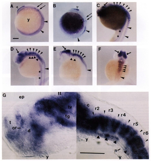
Zash-1b expression in wild-type embryos. 12-hr (A), 14-hr (B), 20-hr (C), 24-hr (D), 30-hr (E, F), and 26-hr (G) embryos were hybridized with a Zash-1b antisense probe. (A-E) Lateral views of whole-mounted embryos. (F) Dorsal view of a 30-hr whole-mounted embryo. (G) Lateral view of a thick longitudinal section of a 26-hr embryo (eyes and lateral head removed) observed by DIC optics (montage of two photographs taken at slightly different planes of focus). Symbols indicating expression sites: open triangles, dorsal telencephalon; thin arrows, early expression sites in dorsal zones of rhombomeres 2 and 4; closed triangles, expression sites in ventral regions of rhombomeres; larger arrowheads, expression in spinal cord; smaller arrowheads, bands of expressing cells in dorsal and intermediate hindbrain; thick short arrows, midbrain expression sites; filled stars, diffuse staining near yolk and gut. Abbreviations are as in the legend to Fig. 3. Scale bars in A and G, 100 μm (A-F at same magnification). |
Reprinted from Developmental Biology, 166, Allende, M.L. and Weinberg, E.S., The expression pattern of two zebrafish achaete-scute homolog (ash) genes is altered in the embryonic brain of the cyclops mutant, 509-530, Copyright (1994) with permission from Elsevier. Full text @ Dev. Biol.