
FIGURE
Fig. 2
- ID
- ZDB-FIG-090515-34
- Publication
- Amsterdam et al., 1995 - The Aequorea victoria green fluorescent protein can be used as a reporter in live zebrafish embryos
- Other Figures
- All Figure Page
- Back to All Figure Page
Fig. 2
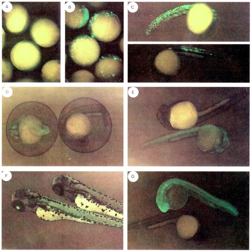
Expression of GFP in injected and transgenic embryos. Injected F0 embryos were observed by epifluorescence microscopy at 5 (B) and 24 hr (C), compared to uninjected embryos (5 hr, A). F1 embryos from lines XIG-3 (D) and XIG-2 (E and F) were observed alongside nontransgenic siblings at 24 (D), 32 (E), and 72 hr (F) postfertilization. An F1 embryo from line XIG-5 was observed alongside a nontransgenic sibling at 24 hr (G). Chorions were removed from the embryos with pronase in A-C, E-G. |
Expression Data
Expression Detail
Antibody Labeling
Phenotype Data
Phenotype Detail
Acknowledgments
This image is the copyrighted work of the attributed author or publisher, and
ZFIN has permission only to display this image to its users.
Additional permissions should be obtained from the applicable author or publisher of the image.
Reprinted from Developmental Biology, 171, Amsterdam, A., Lin, S., and Hopkins, N., The Aequorea victoria green fluorescent protein can be used as a reporter in live zebrafish embryos, 123-129, Copyright (1995) with permission from Elsevier. Full text @ Dev. Biol.