|
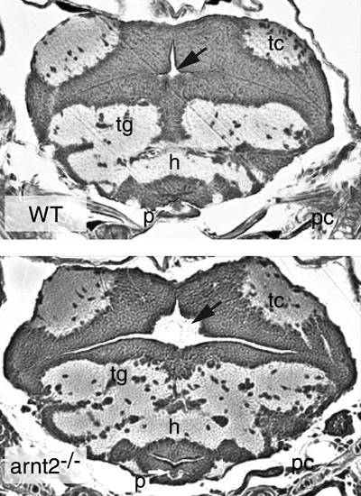
Brain ventricle area is increased in arnt2-/- larvae at 120 hpf. Cross section of the brain of a representative WT larva shows a small ventricle to which a black arrow is pointing (top panel). On the other hand, a cross section taken through the same region of the brain of a arnt2-/- mutant reveals a large brain ventricle (black arrow, bottom panel). The two cross sections were taken at similar locations along the anterior–posterior axis of the brain using the tectum, tc, and presumptive pituitary, p (white arrow), for orientation of the sections. h, Hypothalamus; tg, tegmentum; pc, parachordal cartilage.
|