
Fig. 2
- ID
- ZDB-FIG-090506-46
- Publication
- Alvarez-Delfin et al., 2009 - Tbx2b is required for ultraviolet photoreceptor cell specification during zebrafish retinal development
- Other Figures
- All Figure Page
- Back to All Figure Page
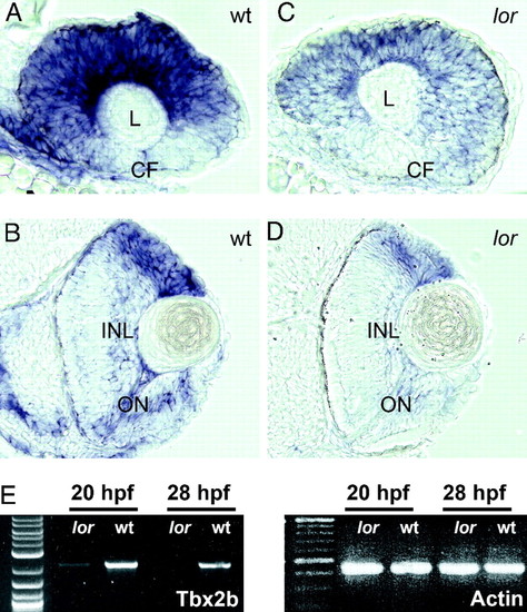
tbx2b expression is reduced in lor mutant embryos. Sagittal sections of WT (A) and lorp25bbtl mutant (C) embryos at 28 hours postfertilization (hpf) following in situ hybridization for tbx2b show labeling throughout the eye, but the lack of expression in the ventral retina near the choroid fissure (CF) and lens (L). Transverse sections of labeled WT (B) and lorp25bbtl embryos (D) at 44 hpf. tbx2b expression is most intense at the dorsal retinal margin and cells adjacent to the developing outer nuclear layer (ONL) as well as the inner nuclear layer (INL) (optic nerve; ON). Note the dramatically reduced expression in the lorp25bbtl embryos. (E) Amplification of tbx2b by RT-PCR from RNA extracted from WT and lorp25bbtl embryos at 20 and 28 hpf demonstrates a reduction in the amount of tbx2b mRNA in the mutant. |
| Gene: | |
|---|---|
| Fish: | |
| Anatomical Terms: | |
| Stage Range: | 20-25 somites to High-pec |