
Fig. 4
- ID
- ZDB-FIG-090506-40
- Publication
- Higashijima et al., 1997 - High-frequency generation of transgenic zebrafish which reliably express GFP in whole muscles or the whole body by using promoters of zebrafish origin
- Other Figures
- All Figure Page
- Back to All Figure Page
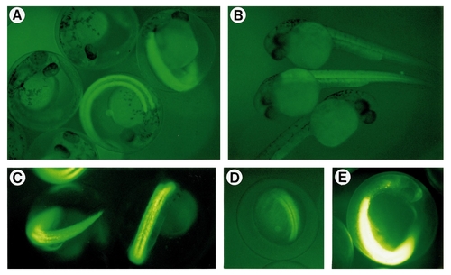
Expression of GFP in transgenic embryos generated using α-actin–GFP. Embryos were viewed through their chorions, except for (B) where chorions were removed. (A) 30-h-old embryos of a B-rank transgenic line. Nonfluorescent embryos are nontransgenic siblings. (B) 28-h-old embryos of C-rank and D-rank transgenic lines. A nontransgenic embryo is also shown. (C) 26-h-old embryos of an A-rank transgenic line. (D) A 13-h-old embryo of an A-rank transgenic line. (E) A 26-h-old embryo of the most fluorescent line (A-rank). Film-exposure time for this figure was much shorter than that in other figures. Due to the high fluorescence of the muscles, other regions of the embryo are clearly visible. |
| Gene: | |
|---|---|
| Fish: | |
| Anatomical Terms: | |
| Stage Range: | 5-9 somites to Prim-15 |
Reprinted from Developmental Biology, 192, Higashijima, S., Okamoto, H., Ueno, N., Hotta, Y., and Eguchi, G., High-frequency generation of transgenic zebrafish which reliably express GFP in whole muscles or the whole body by using promoters of zebrafish origin, 289-299, Copyright (1997) with permission from Elsevier. Full text @ Dev. Biol.