
Fig. 4
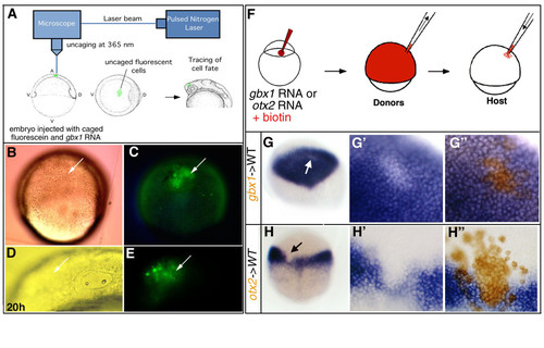
gbx1 induces posterior neural fate via transformation of anterior neural fate and gbx1 and otx2 repress each other cell-autonomously. (A) Schematic drawing of the fluorescein uncaging procedure (for details, see [33]). (B, C) gbx1-injected embryo after uncaging cells in the prospective otx2 domain. (D, E) After 20 h of development the labeled cells come to lie in the anterior of the embryo (arrow). (B, D) Nomarski optics; (C, E) detection of the fluorescent cells (arrow). (F) Experimental procedure for cell transplantations. Donor embryos are generated by co-injecting biotinylated dextran as lineage tracer and gbx1 (300 pg) or otx2 mRNA (300 pg). Cells are taken out of the donor at 40% epiboly and transplanted into a wild-type (WT) host embryo. (G) Chimeric embryo containing cells derived from embryos injected with biotinylated dextran and gbx1mRNA, and stained for otx2 (blue). Unlabeled patch of cells marked by a white arrow. (G′) Close-up of the patch indicated in (G) before biotin staining and (G″) after biotin staining (brown). gbx1 overexpressing cells within the otx2 domain do not express otx2. (H) Chimeric embryos containing cells derived from embryos injected with biotinylated dextran and otx2 mRNA, and stained for gbx1 (blue). Unlabeled patch of cells marked by a black arrow. (H′) Close-up of the patch indicated in (H) before biotin staining and (H″) after biotin staining (brown). otx2 overexpressing cells within the gbx1 domain do not express gbx1. |