
Fig. 6
- ID
- ZDB-FIG-090504-58
- Publication
- Heisenberg et al., 1997 - The function of silberblick in the positioning of the eye anlage in the zebrafish embryo
- Other Figures
- All Figure Page
- Back to All Figure Page
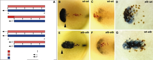
Anterior migration of ventral CNS midline cells is normal at bud stage but impaired at 15 hpf in slb. (A) Schematic diagram showing two different models which might explain the shortening of the ventral CNS midline in slb: (I) reduced anterior migration of the axial mesendoderm (blue) in slb leads to incomplete induction of the ventral CNS midline (hatched, red) in the overlying neural plate (red). (II) Anterior migration of both the axial mesendoderm (blue) and the overlying ventral CNS midline (hatched, red) is impaired in slb (B, E) Position of ventral diencephalic midline cells (brown) in relation to the polster expressing hgg1 (blue) in a wild-type (B) and slb (E) embryo at bud stage. Note that the anterior end of the neural keel (arrowhead) lies to the posterior of the polster in wild type whereas it lies to the anterior of the polster in slb. (C, F) Position of ventral diencephalic midline cells (brown) in relation to the anlage of the optic stalks expressing pax2 (blue) in a wild-type (C) and slb (F) embryo at 15 hpf. (D, G) Transplantation of slb cells (brown) into a wildtype polster expressing hgg1 (blue) (D) and of wild-type cells (brown) into a slb polster expressing hgg1 (blue) (G) do neither phenocopy nor rescue the mutant phenotype. Dorsal views, anterior to the left. |
Reprinted from Developmental Biology, 184(1), Heisenberg, C.P. and Nüsslein-Volhard, C., The function of silberblick in the positioning of the eye anlage in the zebrafish embryo, 85-94, Copyright (1997) with permission from Elsevier. Full text @ Dev. Biol.