
Fig. 5
- ID
- ZDB-FIG-090501-53
- Publication
- Dixon Fox et al., 2009 - Short- and long-range functions of Goosecoid in zebrafish axis formation are independent of Chordin, Noggin 1 and Follistatin-like 1b
- Other Figures
- All Figure Page
- Back to All Figure Page
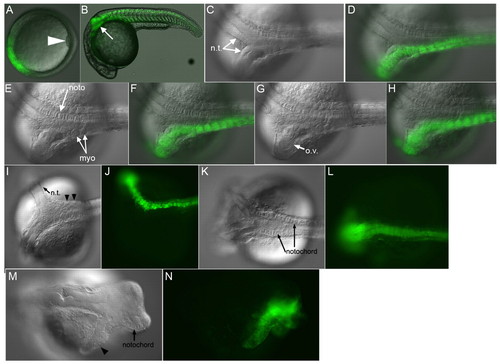
chd induces partial secondary axes. (A-H) Live zebrafish embryo injected with chd RNA. Shield (A), 1 dpf (B-H). (B) Partial secondary axis (arrow, 13/21, 62%). GFP-labeled neural tissue (C,D) (13/13, 100%), myotomes (E,F) (9/13, 69%) and ectopic otic vesicle (G,H) (6/13, 46%). Some injected embryos had beating cardiac tissue (4/13, 31%). (I-L) Live chdtt250 embryos injected ventrally with 48 pg gsc and nog1- and fstl1b-MOs. (I,J) Partial GFP-labeled secondary axis with neural (arrow) and somitic (arrowheads) tissue. (K,L) Secondary axis containing GFP-labeled notochord and neural tissue (3/5, 60%). For an example of an uninjected chd mutant embryo see Fig. 7D. (M,N) Wild-type embryo injected ventrally with chd, fstl1b and nog1 mRNAs. Arrowhead marks neural tissue. ov, otic vesicle. |