
Fig. 2
- ID
- ZDB-FIG-090428-11
- Publication
- Behra et al., 2009 - Phoenix is required for mechanosensory hair cell regeneration in the zebrafish lateral line
- Other Figures
- All Figure Page
- Back to All Figure Page
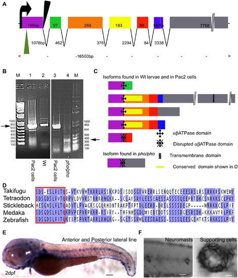
Phoenix is a gene without obvious homologs expressed in the supporting cells of the lateral line. (A) The coding region of the phoenix gene spans approximately 16.5kb with seven spliced exons. Exon 7 is an unusually long ORF of 7758bp. The retroviral integration in the zm allele (green triangle) is in the beginning of the first exon, probably leading to a null allele. In the hi43 allele, the retroviral integration (black triangle) is adjacent to the first splice donor site inducing an aberrant spliced mRNA (see [B] and [C]). The size of exons and introns are indicated. (B) RT-PCR on total RNA extracts of the zebrafish cell line PAC2 (lanes 1 and 3) and 7dpf wild-type (lane 2) and hi43 mutant allele (lane 4) larvae. The expected band of 835bp (left ->) was detected in the PAC2 cell line (lane 1 and 3) and in the wild-type larvae (lane 2). However, in the hi43 mutant lane there is a new 368 bp band representing an aberrant splice event from exon 1 to exon 7 (right ->). (C) We identified five splicing variants from wild-type larvae and the PAC2 cell line and one in the hi43 mutant allele larvae. In the mutant mRNA, the ab ATPase domain (plusses) is disrupted by the aberrant splice (diamonds). A putative transmembrane domain is found in exon 7 (zig-zag bar). The location of the conserved domain described in (D) is shown (yellow rectangle). (D) Alignment that shows the region of homology for putative phoenix genes in four other fish species. One particular stretch seems to be strongly conserved in all fish species, the short SDS- X(3)-SLF-[ILV]-TQ sequence (red box). The remaining sequence for all predicted proteins shows little to no significant identity, beyond enrichment for proline and lysine residues. (E) In situ hybridization with an antisense probe directed against the phoenix gene. At 2dpf the expression was found in all the neuromasts of the anterior and posterior lateral line. (F) Higher magnification of two trunk neuromasts (left panel) and a single neuromast (right panel), showing the typical ring-like staining restricted to the supporting cells. - 100 microns in E, 50 microns in (F, left panel) and 10 microns in (F, right panel). |
| Gene: | |
|---|---|
| Fish: | |
| Anatomical Terms: | |
| Stage Range: | Long-pec to Days 7-13 |