
FIGURE
Fig. 6
- ID
- ZDB-FIG-090415-32
- Publication
- Abdelilah et al., 1997 - Pattern formation in janus-mutant zebrafish embryos
- Other Figures
- All Figure Page
- Back to All Figure Page
Fig. 6
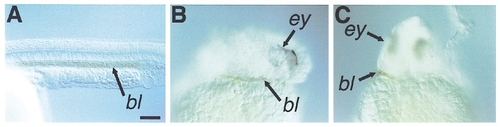
Ventral mesoderm in janus-mutant embryos. (A–C) Diaminofluorene staining of premigratory erythrocytes at the 30-somite stage. (A)Wild-type. (B, C) janus-mutant embryos inwhich the shield was localized to one of the two half-blastoderms have a dorsal half-blastoderm with patches of premigratory erythrozytes. (A, B) Lateral view. (C) Anterior view. bl, erythrozytes; ey, eye. Scale bar is 100 μm. |
Expression Data
Expression Detail
Antibody Labeling
Phenotype Data
Phenotype Detail
Acknowledgments
This image is the copyrighted work of the attributed author or publisher, and
ZFIN has permission only to display this image to its users.
Additional permissions should be obtained from the applicable author or publisher of the image.
Reprinted from Developmental Biology, 184(1), Abdelilah, S. and Driever, W., Pattern formation in janus-mutant zebrafish embryos, 70-84, Copyright (1997) with permission from Elsevier. Full text @ Dev. Biol.