
Fig. 4
- ID
- ZDB-FIG-090415-19
- Publication
- Warga et al., 1998 - Spadetail-dependent cell compaction of the dorsal zebrafish blastula
- Other Figures
- All Figure Page
- Back to All Figure Page
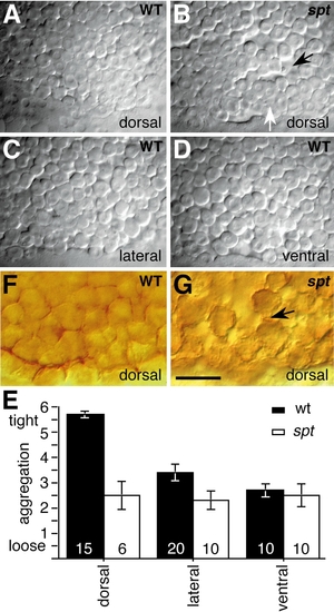
The behavior of blastula marginal cells is changed in spadetail mutants. (A–D) Marginal cells at 40% epiboly, after progression of cell compaction. (E) Assessment of appearance of marginal deep cells just below the surface of the enveloping layer between 30 and 40% epiboly, respective of position in wildtype (solid bar) and spadetail mutants (open bar); lines represent standard error. For reference we ranked A as 6, tightly associated cells, and B and C as 3, tightly and loosely associated cells, and D as 2, loosely associated cells. Cell compaction was statistically compared between wildtype dorsal and lateral cells (Wilcoxon rank sum = 150, n = 15, m = 20) and between wild-type dorsal and mutant dorsal cells (Wilcoxon rank sum = 120, n = 15, m = 6). (F and G) Cell membranes visualized using anti-β-catenin immunostaining at 30% epiboly. Scale bar: 50 μm (A–D), 25 μm (F and G). |
Reprinted from Developmental Biology, 203, Warga, R.M. and Nüsslein-Volhard, C., Spadetail-dependent cell compaction of the dorsal zebrafish blastula, 116-121, Copyright (1998) with permission from Elsevier. Full text @ Dev. Biol.