|
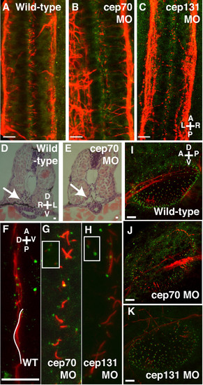
Cilia in the otic vesicle, spine and pronephros at 30 h.p.f. (A-C) Spine of WT, cep70 and cep131 morphants, stained for acetylated tubulin (red, cilia) and gamma tubulin (green, basal bodies). The cilia of the central canal can be seen in between the two nerve tracts. Cilia are shortened in the morphant embryos but basal bodies remain intact. Pronephric ducts – arrows – are enlarged in morphant cep70 embryos (E) compared to WT (D) at 48 h.p.f. . (F-H) Cilia are also reduced in length in the pronephros of the morphants (G, H) compared to WT (F). However, like other tissues, basal bodies are present. Basal bodies are also present in surrounding tissues (inset). One WT cilium has been colored white for clarity. (I-K) Otic vesicles of WT, cep70 and cep131 morphants. Compass in the panels shows orientation: A = anterior, P = posterior, D = dorsal, V = ventral, L = left, R = right. Scale bars: 10 μm.
|