
Fig. 4
- ID
- ZDB-FIG-090408-13
- Publication
- Brösamle et al., 2009 - Nogo-Nogo receptor signalling in PNS axon outgrowth and pathfinding
- Other Figures
- All Figure Page
- Back to All Figure Page
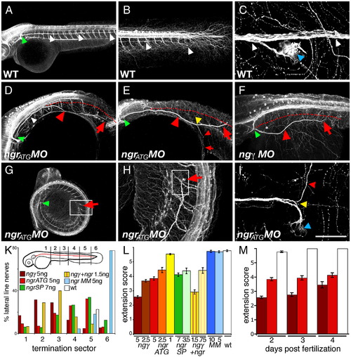
Posterior lateral line (pll) nerve defects result from Nogo-γ and Ngr depletion. (A) Bilaterally, the pll nerve emerges from the pll ganglion (indicated by green arrowhead) and projects caudally along the trunk to reach the ventral base of the tail by the end of 2 dpf (white arrowheads, B). (C) As it traverses the length of the embryo, pll axons innervate sensory organs of the lateral line (blue arrowhead). In (D, E) ngrATG, (F) nogo-γ, and ngrSP (not shown) morphants, the trajectory was frequently abnormal (indicated by red arrowheads), with the pll nerve veering off the correct path (red dotted line), invading inappropriate areas and terminating prematurely (the end of the nerve is indicated by red arrows). (E) In some instances, the pll nerve was found to branch (yellow arrowhead). (G–I) Additionally, many pll nerves terminated prematurely (G–I, H and I higher magnifications of boxed areas in G and H, respectively). NgrATG morphants were frequently curled ventrally (G) and anterior is indicated by (a). (K) The extent of pll outgrowth was scored according to the embryonic sector in which the pll nerve terminated at 48 hpf. In control WT or mismatch morpholino injected larvae, > 95% pll nerves reached sector 5 or 6 (white and blue bars). Following MO injections for ngrATG (red), (green), nogo-γ (brown), or a combination of ngrATG and nogo-γ (striped bars) only between 11% (nogo-γ) and 44% (ngrSP) of nerves terminated in sectors 5 and 6. (L) Average extension scores indicated that pll truncation was dose-dependent and that co-injection of ngr and nogo-γ morpholinos potentiated the effect. (M) Incomplete extension of the pll nerve persisted at 3 and 4 dpf. (A–I) Maximum intensity projections of side view confocal z-series of 2 dpf anti-acetylated tubulin immunostained whole-mount zebrafish larvae. Scale bar A and G 400 μm; B, and D–F 200 μm; H 50 μm; C and I 40 μm. |
| Fish: | |
|---|---|
| Knockdown Reagents: | |
| Observed In: | |
| Stage Range: | Long-pec to Day 4 |
Reprinted from Molecular and cellular neurosciences, 40(4), Brösamle, C., and Halpern, M.E., Nogo-Nogo receptor signalling in PNS axon outgrowth and pathfinding, 401-409, Copyright (2009) with permission from Elsevier. Full text @ Mol. Cell Neurosci.