
Fig. 5
- ID
- ZDB-FIG-090220-46
- Publication
- Hilario et al., 2009 - Semaphorin 5A is a bifunctional axon guidance cue for axial motoneurons in vivo
- Other Figures
- All Figure Page
- Back to All Figure Page
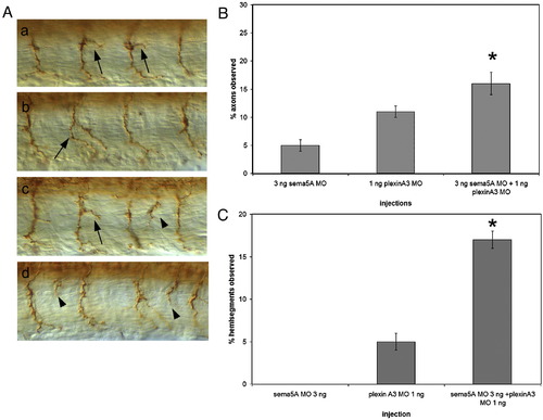
Additive and synergistic interactions of Sema5A and Plexin A3. (A) Axonal phenotypes observed in embryos injected with plexin A3 MO alone or plexin A3 MO and sema5A MO. Arrows indicate moderately and severely branched CaP axons, arrowheads indicate axons exiting the spinal cord at ectopic exit points. (B) Percentage of axons with moderate and severe branching in embryos injected with 3 ng of sema5A MO alone, 1 ng of plexin A3 MO alone and a combination of 3 ng sema5A MO and 1 ng plexin A3 MO. (C) Percentage of hemisegments observed with ectopic axonal exit points in embryos injected with 3 ng sema5A MO, 1 ng plexin A3 MO and a combination of 3 ng sema5A MO and 1 ng plexin A3 MO. Data quantified from 3 ng sema5A MO injections (n = 860 axons, 43 embryos), 1 ng plexin A3 MO injection (n = 2860 axons, 140 embryos) and 3 ng sema5A MO + 1ng plexin A3 MO injections (n = 2760 axons, 128 embryos). Ectopic axonal exit points were not observed in both wild-type uninjected or control MO-injected embryos (not shown). Error bars represent confidence interval for proportions at 95% confidence. Asterisks indicate significant difference between sema5A MO + plexinA3 MO injected and both 3 ng sema5A MO injected and plexinA3 MO injected at p < 0.01 using a Z-test of proportions. |
| Fish: | |
|---|---|
| Knockdown Reagents: | |
| Observed In: | |
| Stage: | Prim-5 |
Reprinted from Developmental Biology, 326(1), Hilario, J., Rodino-Klapac, L.R., Wang, C., and Beattie, C.E., Semaphorin 5A is a bifunctional axon guidance cue for axial motoneurons in vivo, 190-200, Copyright (2009) with permission from Elsevier. Full text @ Dev. Biol.