
Fig. 5
- ID
- ZDB-FIG-090106-12
- Publication
- Jiang et al., 2008 - Exdpf is a key regulator of exocrine pancreas development controlled by retinoic acid and ptf1a in zebrafish
- Other Figures
- All Figure Page
- Back to All Figure Page
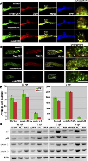
Reduced exdpf Causes Defects of Exocrine Cell Proliferation (A) BrdU labeling result in 33 hpf embryos. Green: MP760:GFP, Red: BrdU staining. Blue: DAPI staining. Dorsal view, anterior to the left. Enlargement represents higher magnification of boxed area in each row. Arrows indicate non-proliferating cells; arrowheads indicate proliferating cells as evidenced by overlapping of red and green colors. (B) BrdU labeling result in 3 dpf embryos. Green: elastase A:GFP, Red: BrdU staining. Lateral view, anterior to the left. Scale bar: 50 μm. Arrows indicate non-proliferating cells; arrowheads indicate proliferating cells as evidenced by overlapping of red and green colors. (C) Quantitative graphs for BrdU incorporation rate in 33 hpf or 3 dpf embryos. The average number of GFP positive cells with BrdU incorporation was obtained by counting BrdU-labeled GFP positive cells from five embryos. Y axis: Mean ± SD. (D) Semiquantitative RT-PCR examination of expression of cyclin D1 and cell cycle inhibitors p21, p27 and cyclin G1 in control (control), exdpf morphants (MO), and exdpf mRNA injected embryos (RNA) at 33 hpf, 2 dpf, 3 dpf, and 5 dpf. In each group, 30 embryos were used to extract total RNA for RT-PCR. |
| Genes: | |
|---|---|
| Antibody: | |
| Fish: | |
| Condition: | |
| Knockdown Reagent: | |
| Anatomical Terms: | |
| Stage Range: | Prim-15 to Day 5 |
| Fish: | |
|---|---|
| Condition: | |
| Knockdown Reagent: | |
| Observed In: | |
| Stage Range: | Prim-15 to Protruding-mouth |