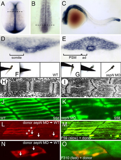
Loss of sepN function disrupts muscle differentiation in the zebrafish embryo. (A–E) sepN is expressed transiently in newly formed somites, adaxial cells (ad), presomitic mesoderm (PSM), and the notochord (ntc). sepN expression in (A and D) 10.5 hpf (two-somite stage), (B and E) 16 hpf, and (C) 24 hpf WT embryos. (A and B), Dorsal views with rostral up; (C) lateral view with rostral left. (D and E) Transverse sections through embryos at the levels indicated in B and C, respectively. (F and G) Three sequential frames illustrate that morphants, unlike WT embryos, rarely move >0.5 cm in response to touch. (H and I) Transmission electron micrographs of fast muscle fibers of 24 hpf (H) WT and (I) sepN morphant embryos indicate disruption of sarcomere organization in morphants. (J and K) Superficial slow muscle fibers of 24-hpf embryos stained with S58 antibody. (L–O) Development of sepN-depleted muscle fibers in WT hosts. sepN-depleted slow (L and M) or fast (N and O) dye-labeled muscle cells (red) were analyzed in mosaic embryos at 24 hpf stained with F59 or F310 antibody (green), respectively. (L and N) Dye-labeled donor cells; (M and O) merged views. *, normal-appearing fibers; arrows, dysmorphic donor fibers.
|
