
Fig. 5
- ID
- ZDB-FIG-081111-34
- Publication
- Mendonsa et al., 1999 - Genetic analysis of tissue interactions required for otic placode induction in the zebrafish
- Other Figures
- All Figure Page
- Back to All Figure Page
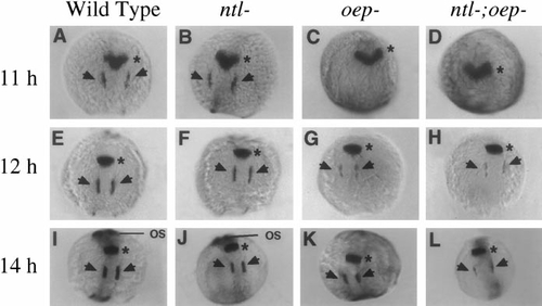
Effects of mesendoderm deficiencies on expression of pax-2.1. Shown are expression patterns of pax-2.1 at (A–D) 11 h, (E–H) 12 h, and (I–L) 14 h. In wild-type (A, E, I) and ntl- (B, F, J) embryos, pax-2.1 is expressed in the primordia of the midbrain–hindbrain border (asterisks) and otic regions (arrows) from 11 h onward, and expression is greatly upregulated in the optic stalks (os) by 14 h. In oep- (C, G, K) and ntl-;oep- embryos (D, H, L), pax-2.1 is expressed normally in the primordium of the midbrain–hindbrain border, but expression in the otic regions is delayed, and staining is not detected in the optic stalks due to disruption of eye development in embryos lacking oep function. |
Reprinted from Developmental Biology, 206, Mendonsa, E.S. and Riley, B.B., Genetic analysis of tissue interactions required for otic placode induction in the zebrafish, 100-112, Copyright (1999) with permission from Elsevier. Full text @ Dev. Biol.