FIGURE

Fig. S4

ID
ZDB-FIG-080925-11
Publication
Del Bene et al., 2008 - Regulation of neurogenesis by interkinetic nuclear migration through an apical-basal notch gradient
Other Figures
All Figure Page
Back to All Figure Page
Fig. S4

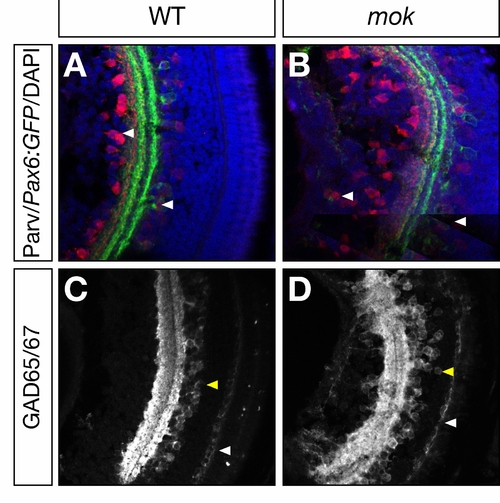
moks309 Retinas Have Normal Number of Amacrine and Horizontal Cells
(A, B) 5 dpf retina section showing pax6:GFP expression (green) and stained with antiparvalbumin antibody (red). Both markers identify a subset of amacrine cells. These cells are specified in normal number in moks309 mutants although their nuclei are sometimes positioned at a greater distance from the inner plexiform layer (IPL) (compare white arrowheads in [A] versus [B]).
(C, D) 5 dpf retina section stained with anti-GAD65/67 antibody. This marker labels both amacrine cells (yellow arrowhead) and horizontal cells (white arrowheads). Their specification is unaffected in moks309 mutants, but amacrine cell nuclei are often mislocalized (compare yellow arrowheads in [C] versus [D]).

Expression Data

Expression Detail
Antibody Labeling
Phenotype Data

Phenotype Detail
Acknowledgments
This image is the copyrighted work of the attributed author or publisher, and ZFIN has permission only to display this image to its users. Additional permissions should be obtained from the applicable author or publisher of the image.

Reprinted from Cell, 134(6), Del Bene, F., Wehman, A.M., Link, B.A., and Baier, H., Regulation of neurogenesis by interkinetic nuclear migration through an apical-basal notch gradient, 1055-1065, Copyright (2008) with permission from Elsevier. Full text @ Cell