
Fig. 2
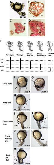
Secondary axis tissues induced following cultured cell implantation. (A) Induced complete secondary axis. (B) Cross section of panel A. The secondary axis had a notochord (no) and neural tube (nt). (C) A second example of induced tissues in the secondary axis in a more posterior region than that shown in panel B. (D) High-magnification view of the region boxed in panel C shows that the secondary axis has a notochord, neural tube, somites (so), and gut (gu). (E) Schematic summary of secondary tissues induced by cell lines. ‘Two eyes’ indicates complete secondary axis with two eyes, trunk and tail; ‘One eye’ means cyclopean with trunk and tail; ‘Trunk with o.v.’ indicates a secondary axis with otic vesicles in the trunk and tail, but without head structures; and ‘Trunk without o.v.’ indicates a secondary axis without head structures or otic vesicle in the trunk, but with a tail. Here, the induced axis was fused with the host endogenous axis in the posterior part of the tail. ‘Part of tail’ indicates a secondary axis only with part of a tail structure. (F, G) Secondary axis with two eyes, trunk and tail induced by ZE6-2 (F) or ZE24-2 (G). (H) Secondary axis with one eye, trunk and tail induced by ZE72-1. (I, J) Secondary axis with trunk, otic vesicle and tail induced by ZE24-2 (I) or ZE120-1 (J). (K, L) Secondary axis with trunk, tail and no otic vesicle induced by ZE24-2 (K) and ZE120-1 (L). (M) Secondary axis with an ectopic tail induced by ZE72-1. Some ectopic tails had floor plate. All embryos were observed at 24 hpf. e, eye of secondary axis; o.v., otic vesicle of secondary axis; t, tail of secondary axis. Arrowheads indicate the implanted DiI-labeled cell clumps at the secondary axis region. Arrows indicate the secondary axis. Scale bar, 100 μm. |
Reprinted from Developmental Biology, 321(2), Hashiguchi, M., Shinya, M., Tokumoto, M., and Sakai, N., Nodal/Bozozok-independent induction of the dorsal organizer by zebrafish cell lines, 387-396, Copyright (2008) with permission from Elsevier. Full text @ Dev. Biol.