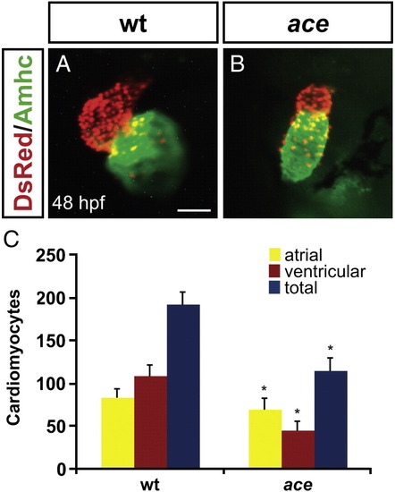
ace mutant embryos have reduced numbers of both ventricular and atrial cardiomyocytes. (A, B) Frontal views of hearts from wild-type (A) and ace mutant (B) embryos at 48 hpf; immunofluorescence detects DsRed (red) in all cardiomyocyte nuclei and atrial myosin heavy chain (Amhc; green) in atrial cells. (A) In a wild-type heart, the ventricle (red) and atrium (green) exhibit typical looping and expansion. (B) In an ace mutant heart, the chambers are unlooped and small, with a particularly apparent reduction of the ventricle. Scale bar represents 50 μm; both images are shown at the same magnification. (C) Quantification of cardiomyocytes at 48 hpf reveals that the numbers of both ventricular and atrial cells are significantly decreased in ace mutants, with ventricular cell number being more affected then atrial cell number. Graph indicates mean and standard deviation for each data set; asterisks indicate statistically significant differences relative to wild-type (p < 0.005, Student's t-test). n = 13 for wild-type, and n = 19 for ace mutants; see also Supplemental Table 1.
|
