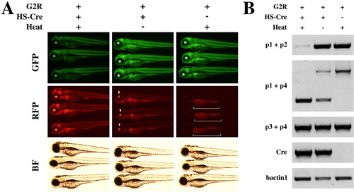
Cre-mediated recombination of G2R in transgenic embryos is detected by fluorescent color change and polymerase chain reaction analysis. A: Fluorescent color conversion by heat induced Cre. Double transgenic embryos (dTg) of HS-Cre and G2R were observed under a dissecting fluorescence microscope 48 hr after the heat shock (left column). The expression of GFP was significantly reduced and RFP was induced, compared to the control embryos of the unheated dTg (center) and G2R single transgenic (sTg, right). The unheated dTg displayed red eyes (arrowheads) due to the spontaneous expression of Cre by the HS promoter in the lens under non-stressed conditions. The sTg retained the strong green signals and showed only the autofluorescent red signals in the yolk (brackets). B: Genomic PCR of recombined G2R. The positions of the primers are represented in Figure 1. Consistent with the color conversion, the non-recombinant-specific bands, p1+p2 and p1+p4 upper, were greatly reduced but the recombinant-specific band, p1+p4 lower, was intensive in heated dTg (left lane). The unheated dTg (center) showed a partial recombination while the sTg (right) showed only the non-recombinant-specific band.
|
