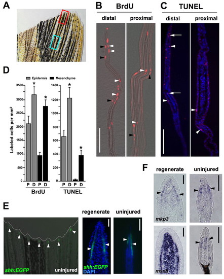
Homeostatic regeneration programs active in zebrafish fins. (A) Ventral lobe of an uninjured caudal fin. The red box indicates areas taken for distal measurements; the blue box is for proximal measures. Each box is 350 μm long, separated by 350 μm, a length chosen because it is the length of a frame at 20x magnification using our imaging equipment. (B) Distal and proximal sections of the caudal fin stained for BrdU incorporation, after a 24-hour labeling period. BrdU-labeled cells (red) are observed in epidermal (black arrowheads) and mesenchymal (white arrowheads) compartments of both distal and proximal regions (distal to top of each image). (C) TUNEL stains of distal and proximal regions, labeling apoptotic cells. TUNEL-positive cells (red) are observed in the epidermal (arrowheads) and mesenchymal (arrows) compartments. Nuclei are labeled with DAPI (blue). (D) Quantification of BrdU and TUNEL labeled cells in epidermal and mesenchymal compartments of proximal (P) and distal (D) fin tissue. (mean±s.e.m.; Student's t-test, *P<0.05). (E) Analysis of a shh:EGFP transgenic reporter strain. (Left) Whole-mount detection of EGFP fluorescence at the distal tips of each ray (arrowheads) of an uninjured shh:EGFP transgenic zebrafish. (Middle and right) shh is expressed in the epidermis adjacent to the blastema in the regenerating fin (arrowheads), and in a similarly restricted epidermal domain at the distal tips of the rays in the uninjured fin (arrowheads). Nuclei are labeled with DAPI (blue). (F) In situ hybridization of tissue sections for mkp3 and msxb. (Top) mkp3 is expressed in the distal lateral epidermis and distal-most mesenchyme of uninjured fins (arrowheads indicate epidermal expression, right), a pattern similar to its expression in the basal epidermal layer and blastema during regeneration (left). (Bottom) msxb expression in the uninjured fin is predominant in distal mesenchyme (right), reminiscent of blastemal expression of msxb after amputation (left). Scale bars: 50 μm.
|
