
Fig. 3
- ID
- ZDB-FIG-080731-9
- Publication
- Parichy et al., 2000 - Mutational analysis of endothelin receptor b1 (rose) during neural crest and pigment pattern development in the zebrafish Danio rerio
- Other Figures
- All Figure Page
- Back to All Figure Page
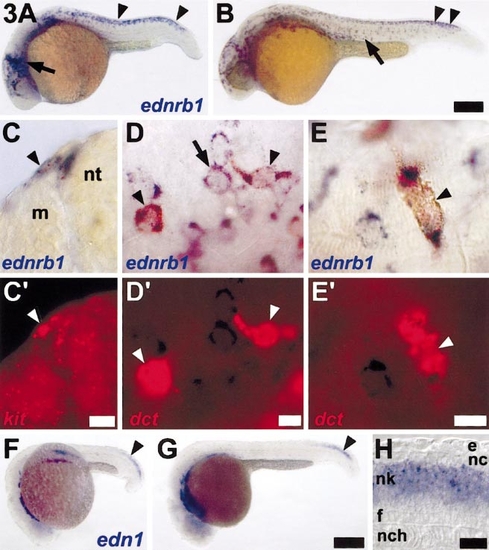
ednrb1 is expressed by cells of the neural crest–melanocyte lineage and is temporally and spatially correlated with endothelin-1 expression in the neural tube. (A) ednrb1+ (blue) cells are abundant over the anterior head, at the midbrain–hindbrain boundary (arrow), within the trunk premigratory neural crest (arrowhead), and in trunk neural crest migratory pathways at 22 h. (B) ednrb1+ cells are more widely scattered over the anterior trunk (arrow) but are still present in the premigratory neural crest in the posterior trunk (arrowhead) at 27 h. (C–E) Two-color in situ hybridization reveals ednrb1 expression by melanocytes and their precursors. (C,C′) Corresponding brightfield and epifluorescence views revealing ednrb1+ cells (blue, C) adjacent to the neural tube (nt) at the entrance to dorsolateral (arrow head) and ventrolateral neural crest migratory pathways. Both cells coexpress the melanocyte lineage marker, kit (red, C′). Shown is a 12-μm cryosection through the posterior trunk of a wild-type embryo. (D,D′) Corresponding images of a whole mount 24-h embyro showing cells coexpressing ednrb1+ (blue, D) and dct (red, D′; arrowheads) near the midbrain–hindbrain boundary. Adjacent cells express only ednrb1 (e.g., arrow). (E,E′) During later development (here, 28 h), ednrb1 continues to be expressed (blue, E) by lightly melanized melanocytes (arrowhead) that coexpress dct (red, E′). (F–H) endothelin-1 is expressed in the posterior trunk and tail developing neural tube. (F) endothelin-1 (edn1) expression is evident in the neural keel posteriorly (arrowhead) as well as in lateral mesoderm and in the vicinity of branchial arches anteriorly, at 20 h. (G) endothelin-1 continues to be expressed in the neural keel of the developing post-anal tail (arrowhead), here shown at 24 h. (H) A longitudinal optical section through the midline posterior trunk of a whole mount 22-h embryo reveals endothelin-1 expression within the middle of the neural keel (nk), although not in the floorplate (f) or dorsal-most layer of neural keel cells (presumptive neural crest, nc). e, epidermis; nch, notochord. Scale bars: (A,B) 400 μm; (C,C′) 10 μm; (D,D′) 10 μm; (E,E′) 10 μm; (F,G) 70 mm; (H) 20 μm. |
Reprinted from Developmental Biology, 227(2), Parichy, D.M., Mellgren, E.M., Rawls, J.F., Lopes, S.S., Kelsh, R.N., and Johnson, S.L., Mutational analysis of endothelin receptor b1 (rose) during neural crest and pigment pattern development in the zebrafish Danio rerio, 294-306, Copyright (2000) with permission from Elsevier. Full text @ Dev. Biol.