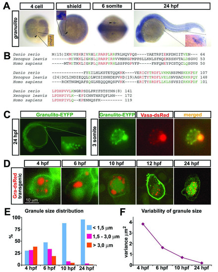
Expression pattern of granulito RNA and subcellular localization of the Ganulito protein. A) Whole-mount in situ hybridizations using granulito antisense RNA probe at the indicated stages. granulito is enriched at the region where the germ plasm resides (cleavage furrows, arrowheads) and is expressed in the primordial germ cells at later stages (arrowheads). B) Alignment of the zebrafish Granulito protein with those from Xenopus leavis and Homo sapiens. Red signifies conservation in all 3 species, green labels conserved substitutions. C) Subcellular localization of Granulito-EYFP. Granulito localizes specifically to germ cell granules as it colocalizes with Vasa protein. D) Germ cells of kop-granulito-dsRedex-nos1-3′UTR transgenic fish whose membrane and nucleus are labeled in green (except for the 4 hours stage where the nucleus is not labeled). Initially, germ cell granules with a large variety of sizes are observed. As development proceeds, the variation in germ cell granule size decreases. E) Granule size distribution at different stages. F) Variance of granule size at different stages. N >100 granules for each time point.
|
