
Fig. 3
- ID
- ZDB-FIG-080620-7
- Publication
- Alexandre et al., 1999 - Somatotopy of the lateral line projection in larval zebrafish
- Other Figures
- All Figure Page
- Back to All Figure Page
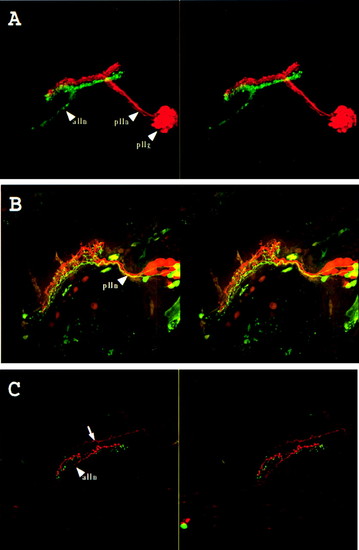
Double labeling of the lateral line system. (A) Labeling of the midbody posterior lateral line nerve (plln) with rhodamine-dextran (red) and of the anterior lateral line nerve (alln) with fluoresceine-dextran (green). Stereo pair of a side view seen from the outside of the animal. Anterior is to the left and dorsal to the top. pllg, posterior lateral line ganglion. The anterior lateral line ganglion could not be imaged because of the limited working distance of the lens. (B) Labeling of the afferents of the second neuromast of the midbody posterior lateral line (2 in Fig. 1) with fluoresceine-dextran and the last two neuromasts of the same line (1 in Fig. 1) with rhodamine-dextran. Stereo pair of a dorsolateral view seen from the outside of the animal. Anterior is to the left and dorsal to the top. plln, posterior lateral line nerve. (C) Labeling of the afferents of a neuromast located dorso anteriorly to the ear (3 in Fig. 1) with rhodamine-dextran and the last neuromast of the supra orbital line (4 in Fig. 1) with fluoresceine-dextran. Stereo pair of a dorsolateral view seen from the outside of the animal. Anterior is to the left and dorsal to the top. alln, anterior lateral line nerve. The arrow indicates trigeminal descending fibers, which often were labeled in these preparations. Magnification: x160. |