
Fig. 5
- ID
- ZDB-FIG-080617-10
- Publication
- Anderson et al., 2008 - Loss of unc45a precipitates arteriovenous shunting in the aortic arches
- Other Figures
- All Figure Page
- Back to All Figure Page
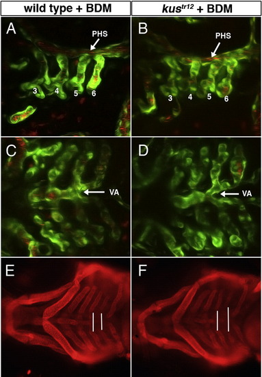
Lack of blood flow can rescue AVM formation in kustr12 mutants. Ninety-eight percent (n = 117) of wild type or heterozygous embryos treated with 2,3-butanedione monoxime (BDM; stops heart beat) between 47 and 53 hpf exhibited normal aortic arches (A), ventral aorta (C), and jaw and pharyngeal cartilages (E). This treatment rescued aortic arch (B) and ventral aorta (D) phenotypes in 67% of kustr12 mutants (n = 29), but had no effect on the basibranchial cartilage phenotype (vertical lines in Panel F). A–D: 2D confocal projections of 74 hpf TG(flk1:GFPla116;gata1:dsRed) embryos, labeling endothelial and blood cells green and red, respectively. G–H: Macro images of 4 dpf embryos stained with anti-collagen II. A, B: lateral views, anterior the left. C–F: ventral views, anterior to the left. PHS, primary head sinus. VA, ventral aorta. |
| Genes: | |
|---|---|
| Antibody: | |
| Fish: | |
| Condition: | |
| Anatomical Terms: | |
| Stage Range: | Protruding-mouth to Day 4 |
| Fish: | |
|---|---|
| Condition: | |
| Observed In: | |
| Stage Range: | Protruding-mouth to Day 4 |
Reprinted from Developmental Biology, 318(2), Anderson, M.J., Pham, V.N., Vogel, A.M., Weinstein, B.M., and Roman, B.L., Loss of unc45a precipitates arteriovenous shunting in the aortic arches, 258-267, Copyright (2008) with permission from Elsevier. Full text @ Dev. Biol.