|
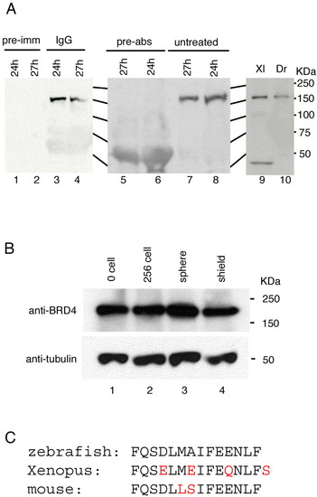
Brd4 protein analysis. A: Antibody characterization. Polyclonal antibody against the Brd4 C-terminal 14 residues detected an approximately 150-kDa protein in zebrafish embryonic extracts (A, lanes 3, 4, 7, 8, and 10). Preimmune serum (pre-imm, lanes 1 and 2) did not detect this protein. Antibody preincubated with antigen peptide (pre-abs, lane 5 and 6) failed to detect the 150-kDa protein. Anti-Brd4 antibody cross-reacts with a protein of predicted size in extracts from Xenopus embryos (lane 9). B: Brd4 protein during zebrafish embryogenesis. Extracts equivalent to two embryos were loaded in each lane. Lower panel shows α-tubulin as a loading control. Stages are indicated for each lane. C: The C-terminal amino acid sequence of zebrafish, Xenopus, and mouse is shown with differences in red.
|