
Fig. 1
- ID
- ZDB-FIG-080606-1
- Publication
- Ma et al., 2001 - Production of zebrafish germ-line chimeras from embryo cell cultures
- Other Figures
- All Figure Page
- Back to All Figure Page
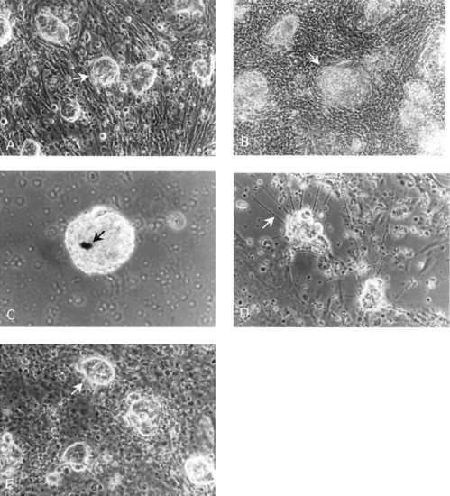
Phase contrast photomicrographs of zebrafish embryo cell cultures. (a) Culture (24 h) of embryo cell aggregates (arrow) on a feeder layer of RTS34st cells. (b) Culture (20 day old) of embryo cell aggregates (arrow) grown on an RTS34st feeder layer. (c) Culture (15 day old) maintained in the absence of feeder cells showing the presence of melanocytes (arrow). (d) Culture (10 day old) maintained in the absence of feeder cells showing the presence of neurites (arrow), indicating that neural cell differentiation has occurred. The neurites begin to appear in the culture on approximately day 5. (e) Culture (25 day old) of embryo cells in RTS34st cell-conditioned medium without a feeder layer, illustrating embryo cell aggregates (arrow) on a monolayer of embryo fibroblasts. |