
FIGURE
Fig. 1
- ID
- ZDB-FIG-080604-57
- Publication
- Zhang et al., 2008 - Cell cycle progression is required for zebrafish somite morphogenesis but not segmentation clock function
- Other Figures
- All Figure Page
- Back to All Figure Page
Fig. 1
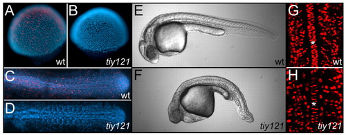
tiy121 mutants have a cell cycle defect. (A-D) Phosphorylated Histone H3 staining (PHH3, red) marks mitotic cells in wild-type embryos at the shield (A) and 10-somite stage (C). No mitotic nuclei are seen in tiy121 embryos at the shield (B) or 10-somite stage (D). Nuclei are stained with DAPI (blue). (E,F) Wild-type (E) and tiy121-/- (F) embryos at 30 hpf. (G,H) BrdU labeling (red) in the trunks of wild type (G) and tiy121-/- (H) at the 14-somite stage. Asterisks label the notochord. BrdU was injected into the yolk at the 8-somite stage. In C-F, anterior is left; in G and H, anterior is up. |
Expression Data
Expression Detail
Antibody Labeling
Phenotype Data
| Fish: | |
|---|---|
| Observed In: | |
| Stage Range: | Shield to Protruding-mouth |
Phenotype Detail
Acknowledgments
This image is the copyrighted work of the attributed author or publisher, and
ZFIN has permission only to display this image to its users.
Additional permissions should be obtained from the applicable author or publisher of the image.
Full text @ Development