
Fig. 1
- ID
- ZDB-FIG-080522-11
- Publication
- Chin et al., 2000 - Heart and gut chiralities are controlled independently from initial heart position in the developing zebrafish
- Other Figures
- All Figure Page
- Back to All Figure Page
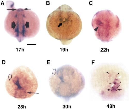
Heart position and heart tube looping direction in the developing zebrafish. Whole-mount in situ hybridization with BMP4 alone. Embryos, at the h.p.f. indicated, are viewed dorsally, with rostral to the top. Bar 200 μm. (A) Heart primordia are present in the lateral plate mesoderm (thick arrows) at 17 h.p.f. Thin arrows denote olfactory regions. (B) At the onset of heart tube assembly at 19 h.p.f., BMP4-expressing cells form a shallow cylinder (arrow), oriented parallel to the dorsal–ventral axis. (C) By 22 h.p.f., the heart tube cylinder (arrowhead) has become oriented in a nearly left–right direction. (D, E) At 28–30 h.p.f., the rostral end of the heart tube (open arrows) continues to migrate back toward the midline. Closed arrow denotes visceral arch. (F) By 48 h.p.f., the heart tube (outlined by the dots) has clearly looped, with the ventricle (portion between the white arrows and the black arrows) lying to the right of the atrium (portion between arrowheads and white arrows). |
Reprinted from Developmental Biology, 227(2), Chin, A.J., Tsang, M., and Weinberg, E.S., Heart and gut chiralities are controlled independently from initial heart position in the developing zebrafish, 403-421, Copyright (2000) with permission from Elsevier. Full text @ Dev. Biol.