
Fig. 6
- ID
- ZDB-FIG-080508-38
- Publication
- Henry et al., 2001 - Roles for zebrafish focal adhesion kinase in notochord and somite morphogenesis
- Other Figures
- All Figure Page
- Back to All Figure Page
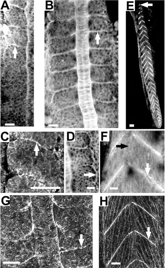
Fak protein and phosphorylated Fak become concentrated at somite boundaries during somitogenesis and persist throughout myotome development. (A–D, G) Dorsal views. (E, F, H) Side views. (A–F) Fak stained with anti-human Fak. (G, H) Phospho-Fak stained with anti-pY397FAK. (A) 10-somite embryo (white arrow designates last formed somite). (B) 7-somite embryo. White arrow designates a somite boundary. (C) A higher magnification of Fak in a 10-somite embryo. Arrow indicates the basal side of border cells that abut the intersomitic furrow (arrow). (D) 7-somite embryo. Arrow indicates the lateral edges of the lateral epithelial cells of a somite. (E, F) 32-h embryos. (G) 10-somite embryo. (H) 30-h embryo. Scale bars, 20 μm, with the exception of (E), where scale bar is 40 μm. |
Reprinted from Developmental Biology, 240(2), Henry, C.A., Crawford, B.D., Yan, Y.-L., Postlethwait, J., Cooper, M.S., and Hille, M.B., Roles for zebrafish focal adhesion kinase in notochord and somite morphogenesis, 474-487, Copyright (2001) with permission from Elsevier. Full text @ Dev. Biol.