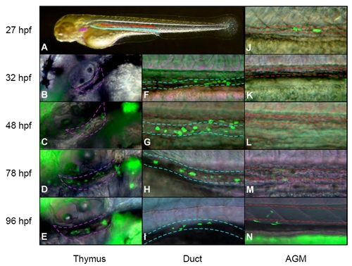
A cmyb:eGFP transgene marks cells in the AGM, along the pronephric ducts and in the thymic lobes. (A) Overview of regions shown in high-magnification fluorescent images. Purple region denotes left thymic lobe, blue region the left pronephric duct and red region the AGM (space between axial vessels). (B-E) cmyb is expressed in the first thymic immigrants beginning at ∼48 hpf. (F-I) GFP+ cells appear along the pronephric tubules beginning at ∼32 hpf, and increase in number over time. Broken lines indicate the boundaries of the duct. (J-N) Within the embryo, GFP+ cells are first observed in the AGM region at ∼27 hpf. After 48 hpf, the AGM region greatly expands as the aorta and vein move apart. The upper broken line indicates the ventral wall of the dorsal aorta, the lower line indicates the dorsal wall of the cardinal vein. GFP+ ductal cells appear ventrolateral to cells within the demarcated AGM region. Images are merged fluorescence and Nomarski photographs. Embryos are positioned anterior towards the left and dorsal side upwards.
|
