
Fig. 2
- ID
- ZDB-FIG-080502-34
- Publication
- Möller et al., 2008 - Expression of trpC1 and trpC6 orthologs in zebrafish
- Other Figures
- All Figure Page
- Back to All Figure Page
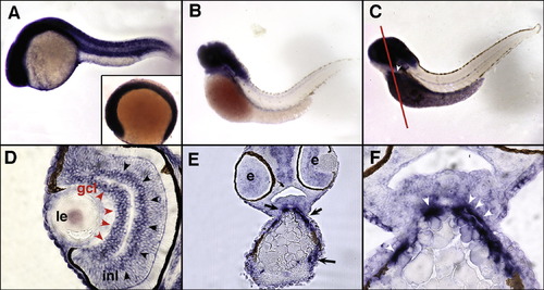
Expression of zebrafish trpC1 by whole mount in situ hybridization and histological analysis. Expression of trpC1 mRNA is ubiquitous in six somite embryos (A; inset) and stages up to and including 24 hpf (A). At 56 hpf, expression is restricted to the head with no detectable expression in the trunk. (B) Strong head expression of trpC1 persists until 72 hpf (C), in addition to expression in the outflow tract of the heart (white arrowhead; white line denotes plane of section in panels E and F). Histological examination of 72 hpf embryos reveals specific expression of trpC1 in the ganglion cell layer of the eye (gcl, red arrowheads) and in the inner nuclear layer (inl, black arrowheads). Anterior sections of 72 hpf embryos (line in C) confirm expression of trpC1 in the outflow track (E, black arrows). A magnified view (F) shows a high level of expression in the cells associated with the outflow tract (white arrowheads). Le, lens; e, eye; gcl, ganglion cell layer; inl, inner nuclear layer. |
| Gene: | |
|---|---|
| Fish: | |
| Anatomical Terms: | |
| Stage Range: | 5-9 somites to Protruding-mouth |
Reprinted from Gene expression patterns : GEP, 8(5), Möller, C.C., Mangos, S., Drummond, I.A., and Reiser, J., Expression of trpC1 and trpC6 orthologs in zebrafish, 291-296, Copyright (2008) with permission from Elsevier. Full text @ Gene Expr. Patterns