|
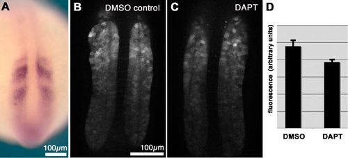
Effect of Loss of Notch Signalling on the Expression of a GFP Reporter for her1
(A) ISH staining for gfp mRNA in a transgenic embryo shows a pattern of expression similar to the normal her1 expression pattern.
(B) GFP fluorescence of a transgenic embryo in the living state treated with DMSO (control), as seen by confocal microscopy in an optical section. Note that the magnification is higher than in (A), and the optical section shows only the PSM.
(C) Corresponding embryo treated with DAPT and imaged in the same way as in (B).
(D) Measured average fluorescence intensities in the PSM of transgenic embryos containing the reporter, after treatment with either 100 μM DAPT or DMSO (control) medium. Treatment was from 5.5 hpf up to 16-somite stage. Fluorescence levels were lower in the DAPT-treated embryos than in the controls by a factor of 0.81 ± 0.07 (mean ± SEM, n > 12 embryos of each type). Error bars show standard error of the mean.
|