
Fig. 5
- ID
- ZDB-FIG-080429-23
- Publication
- Murciano et al., 2002 - Ray-interray interactions during fin regeneration of Danio rerio
- Other Figures
- All Figure Page
- Back to All Figure Page
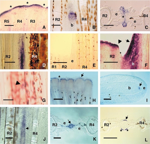
(A) Normal expression pattern of msxD in the epidermis distal to the blastema in control fins. Note that msxD is expressed in the epidermis covering the ray and the interray (asterisks). (B) Following single ray ablation, msxD expression is restricted to the epidermis overlying the distal blastema of the regenerating ray. (C) Cross-section of a fin similar to that shown in (B) at the level of the ray blastema, showing msxD expression in the epithelial tissue surrounding the blastema (asterisk). The lateral epidermis (e) does not express this gene at this level. (D) msxA expression is similar to that of msxD under the same experimental conditions. (E) When ablation of R3 is preceded by the amputation of R1 and R2 a few days before, the epidermis covering R3 (e) remains in contact with neighboring R2 blastema. (F). Expression of msxD under this experimental condition (h) when the ray blastema has reached the distal epidermis (arrow). Regeneration of tissue without ray is faster than normal regeneration but stops at the level of the neighboring blastemas (arrowhead) because it needs two neighboring rays to occur. Control neighboring ray blastemas do not induce msxD expression in the interray (asterisk). (G) During growth of the R1 grafted piece to the I9 interray, msxD expression in the overlying epidermis of the host is only induced at the level of the graft blastema (arrowhead). (H) During normal regeneration, bmp4 is expressed in the distal part of the regenerate (arrow). No expression is observed in the interray region (asterisk). (I) Longitudinal sections of ray shows that bmp4 expression is restricted to the distal blastema (arrow). (J) During single ray regeneration, bmp4 expression is induced in the region occupied by the distal ray blastema (arrow). (K) Cross-section of a single ray regenerate showing that bmp4 is expressed in the distal ray blastema (arrow). There is no expression in the interray blastema and covering epidermis (e). (L) During single ray regeneration, shh is expressed in the basal layer of the epidermis covering the proximal blastema (arrow). Note that shh is expressed in two subdomains (asterisks) in one of the hemirays, suggesting that a bifurcation is about to occur. Scale bars represent 25 (I), 50 (C, K), 100 (A–B, D, F–G, J, L), 200 (E), and 250 μm (H). R1, R2, R3, R4, and R5, original first, second, third, fourth, and fifth ray, respectively; e, epidermis; b, ray blastema. |
Reprinted from Developmental Biology, 252(2), Murciano,C., Fernández, T.D., Durán, I., Maseda, D., Riuz-Sánchez, J., Becerra, J., Akimenko, M.-A., and Marí-Beffa, M., Ray-interray interactions during fin regeneration of Danio rerio, 4-224, Copyright (2002) with permission from Elsevier. Full text @ Dev. Biol.