
Fig. 5
- ID
- ZDB-FIG-080424-81
- Publication
- Lawson et al., 2002 - In vivo imaging of embryonic vascular development using transgenic zebrafish
- Other Figures
- All Figure Page
- Back to All Figure Page
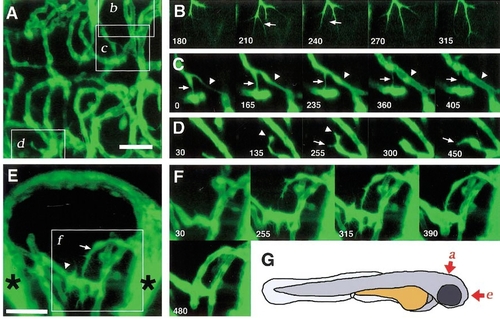
Time-lapse analyses of cranial vessels in TG(fli1:EGFP)y1; albb4/b4 larvae using multiphoton imaging. (A) Central arteries in zebrafish hindbrain at approximately 3.5 dpf, dorsal view, anterior to the right; see (G) for orientation of images. (B–D) Time-lapse sequences of the regions delineated by the corresponding boxes (b, c, d) in (A). (B) A growing central artery exhibiting extension and regression of numerous filopodial extensions (arrows in 210′ and 240′; see Movie 2). (C) A branched central artery exhibits regression of one connection (indicated by an arrow throughout the sequence), while the other connection is retained and appears lumenized by the end of the sequence (arrowhead; see Movie 3). (D) A central artery sprout (arrowhead in time points 135′ and 255′) contacts a potential target vessel via its numerous filopodia. It then regresses and sprouts in a different direction (arrow, time points 255′ and 450′; see Movie 4). (E) Zebrafish forebrain at approximately 2.5 dpf showing communicating vessel (arrow) and palatocerebral artery (arrowhead), frontal view, dorsal up; see (G) for orientation. Brightly labeled plexuses of vessels behind the eye are noted (asterisks). Boxes indicate contact with the palatocerebral artery. Both the communicating vessel and palatocerebral artery exhibit filopodial extensions on the surface (for example, see time point 255′). Filopodial activity in both vessels diminishes just prior to lumenization of the communicating vessel (compare time points 315′ and 480′; see Movie 5). Time-lapse movies and selected three-dimensional reconstructions are available at http://zfish.nichd.nih.gov/zfatlas/fli-gfp/Fli_Home.html. Scale bar, (A, E) 50 μm. |
Reprinted from Developmental Biology, 248(2), Lawson, N.D. and Weinstein, B.M., In vivo imaging of embryonic vascular development using transgenic zebrafish, 307-318, Copyright (2002) with permission from Elsevier. Full text @ Dev. Biol.