
Fig. 9
- ID
- ZDB-FIG-080423-22
- Publication
- Parichy et al., 2003 - Essential role for puma in development of postembryonic neural crest-derived cell lineages in zebrafish
- Other Figures
- All Figure Page
- Back to All Figure Page
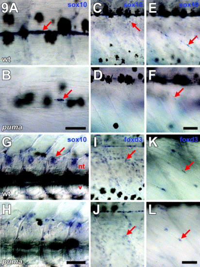
puma promotes development of sox10-expressing cells but is not essential for normal foxd3 expression during metamorphosis. All images are side views of whole-mount larvae during middle stages of pigment pattern metamorphosis with anterior to the left. (A–H) mRNA in situ hybridization for sox10. (I–L) In situ hybridization for foxd3. (A, C, E, G, I, K) Wild-type larvae. (B, D, F, H, J, L) puma mutant larvae. (A) In wild-type larvae, sox10+ presumptive glia are abundant around the posterior trunk lateral line nerve (arrow). Melanophores can be seen in the skin, above the plane of focus. (B) In puma mutants, a severe reduction in sox10+ cells is evident particularly along the posterior trunk lateral line nerve. Only a few sox10+ cell are present at this location initially (arrow), though this number increases during later development. (C, E) Lower magnification views showing the flank of a wild-type larva, either superficially just beneath the skin (C), or deeper within the myotomes (E). Scattered sox10+ cells are evident in both locations, as well as lining the posterior trunk lateral line nerve (near the tops of the panels). (D, F) Corresponding views of an equivalent stage puma mutant larva showing the near complete absence of sox10+ cells both superficially and internally. A single sox10+ within the myotome is indicated (F, arrow). (G, H) Deeper views, just lateral to the larval midline. (G) In wild-type larvae, sox10+ presumptive glia are present in dorsal root ganglia (arrow), adjacent to the neural tube (nt). v, vertebrae. A few melanophores are present dorsal and ventral to the neural tube. (H) In puma mutants, sox10+ cells are present medially but appear relatively disorganized as compared with wild-type larvae. (I) In wild-type larvae, foxd3+ cells are widely distributed and are especially abundant within the myotomes (arrow). Some foxd3+ cells also are present around the posterior lateral line nerve, though these cells are fewer in number compared with sox10+ cells at this location. (J) In puma mutants, similar numbers of foxd3+ cells (arrow) are present compared with wild-type. (A, B) 60 μm; (C–F, I, J) 80 μm; (G, H) 80 μm; (K, L) 40 μm. |
Reprinted from Developmental Biology, 256(2), Parichy, D.M., Turner, J.M., and Parker, N.B., Essential role for puma in development of postembryonic neural crest-derived cell lineages in zebrafish, 221-241, Copyright (2003) with permission from Elsevier. Full text @ Dev. Biol.