
Fig. 2
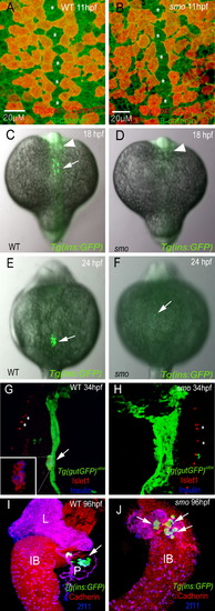
Absence of Dorsal Pancreatic Bud Derived β Cells in smo Mutants (A and B) Ventral confocal images of Tg(sox17:DsRed)s903 (red) and β-catenin (green) expression, comparing wild-type (A) and smo mutants (B) at 18 hpf (mid-trunk region). Tg(sox17:DsRed)s903-expressing endodermal cells show a similar arrangement and distribution around the midline (asterisks) in wild-type (A) and smo mutants (B). (C–F) Dorsal views of Tg(ins:GFP) embryos stained for GFP (green), comparing wild-type (C and E) and smo mutants (D and F) at 18 (C and D) and 24 (E and F) hpf. (C and D) Tg(ins:GFP)-expressing cells are completely missing in 18 hpf smo mutants. Cells in the anterior neural tube (arrowhead) also express the ins:GFP transgene. (E) Tg(ins:GFP)-expressing cells form a cluster in 24 hpf wild-type embryos (arrow). (F) Tg(ins:GFP)-expressing cells are completely missing in most smo mutants, though in approximately 5% of the mutants (n > 100 mutants examined), one or two Tg(ins:GFP)-expressing cells can be observed ([F], arrow). (G–J) Confocal projections of wild-type (G and I) and smo mutant (H and J) endoderm at 34 (G and H) and 96 (I and J) hpf. (G and H) Tg(gutGFP) s854 embryos were stained for Islet1 (red) and Insulin (blue). (G) The endodermal cells (green) form a solid multicellular rod, and pancreatic endocrine cells (arrow and inset) aggregate into one cluster in wild-type embryos. (H) In smo mutants, the endodermal cells fail to condense into a midline rod, and pancreatic endocrine cells are completely absent. (I and J) Tg(ins:GFP) embryos were stained for pan-Cadherin (red) and 2F11 (blue) to mark the endoderm and ductal structures, respectively. (I) Tg(ins:GFP)-expressing β cells (arrow) form a cluster inside the wild-type pancreas. (J) Although the morphology of the pancreas and liver is disrupted, Tg(ins:GFP)-expressing β cells (arrows) appear in smo mutants in ventral pancreatic bud derived tissues. L, liver; P, pancreas; IB, intestinal bulb. |
| Genes: | |
|---|---|
| Fish: | |
| Anatomical Terms: | |
| Stage Range: | 1-4 somites to Day 4 |
| Fish: | |
|---|---|
| Observed In: | |
| Stage: | Day 4 |
Reprinted from Developmental Cell, 14(4), Chung, W.S., and Stainier, D.Y., Intra-endodermal interactions are required for pancreatic beta cell induction, 582-593, Copyright (2008) with permission from Elsevier. Full text @ Dev. Cell