
Fig. 4
- ID
- ZDB-FIG-080418-19
- Publication
- Hsiao et al., 2003 - Transgenic zebrafish with fluorescent germ cell: a useful tool to visualize germ cell proliferation and juvenile hermaphroditism in vivo
- Other Figures
- All Figure Page
- Back to All Figure Page
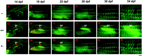
Visualization of juvenile hermaphroditism in living TG(β-actin:EGFP) by following the fluorescent appearance of gonads. (A–F) In -- group juveniles (33%), no fluorescent appearance of gonads could be detected from juveniles throughout sexual maturation. (G–L) In ++ group juveniles (44%), gonads were fluorescent from juveniles and continued to fluoresce throughout sexual maturation. (M–R) In +- group juveniles (23%), gonads transiently fluoresced at the juvenile stage but gradually lost their fluorescence in later development stages. Fish in all pictures are viewed laterally and the anterior is to the left. All pictures were recorded at the same exposure time (2 s). Development stages are indicated in each panel. dpf, day postfertilization; gc, germ cells. Scale BAR = 500 μm in (A, G, M) and 1 mm in all other pictures. |
Reprinted from Developmental Biology, 262, Hsiao, C.-D. and Tsai, H.-J., Transgenic zebrafish with fluorescent germ cell: a useful tool to visualize germ cell proliferation and juvenile hermaphroditism in vivo, 313-323, Copyright (2003) with permission from Elsevier. Full text @ Dev. Biol.