
Fig. 5
- ID
- ZDB-FIG-080418-10
- Publication
- Griffin et al., 2003 - Interplay between FGF, one-eyed pinhead, and T-box transcription factors during zebrafish posterior development
- Other Figures
- All Figure Page
- Back to All Figure Page
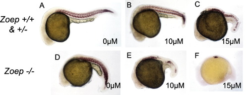
The Zoep mutant phenotype is enhanced by FGFR inhibition. Embryos at 24 h of development, anterior to left, hybridized to detect myoD expression (brown stain). (A–C) Zoep +/?; (D–F) Zoep -/-. Defects in wild-type tail somitic mesoderm are first observed at 15 μM (C), as described in Fig. 3. (D) Untreated Zoep mutant embryo. (E) Zoep mutant embryos treated with 10 μM show aberrations in the number of myoD-positive cells. Unlike the defects observed in wild-type embryos treated with 10 μM SU5402, loss of muscle did not occur strictly from posterior to anterior, as indicated by the gap in myoD expression in the posterior trunk. Muscle staining in the tail was continuous across the midline, indicating the absence of posterior notochord. (F) Zoep mutant embryos treated with 15 μM; myoD staining is only detected in the anterior trunk. |
Reprinted from Developmental Biology, 264(2), Griffin, K.J. and Kimelman, D., Interplay between FGF, one-eyed pinhead, and T-box transcription factors during zebrafish posterior development, 456-466, Copyright (2003) with permission from Elsevier. Full text @ Dev. Biol.