
Fig. 5
- ID
- ZDB-FIG-080415-9
- Publication
- Pei et al., 2008 - Identification and characterization of a novel gene differentially expressed in zebrafish cross-subfamily cloned embryos
- Other Figures
- All Figure Page
- Back to All Figure Page
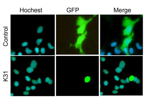
Sub-cellular localization of pEGFP-K31 expressed in EPC cells. The sub-cellular localization of control (pEGFP-N3) and pEGFP-K31 expressed GFP signals in EPC cells was in the upper and lower rows, respectively. Therein, blue signals represented the cell nuclei stained by Hochest 33342; Green signals represented the expression of pEGFP-N3 and pEGFP-K31 fluorescence proteins in EPC cells, respectively; Merge represented overlapping the images of pEGFP-N3 or pEGFP-K31 fluorescent protein with the images of cell nuclei stained by Hochest 33342. All three panels had the same view field at 24 h after transfection. |