
Fig. 7
- ID
- ZDB-FIG-080411-24
- Publication
- Insinna et al., 2008 - The homodimeric kinesin, Kif17, is essential for vertebrate photoreceptor sensory outer segment development
- Other Figures
- All Figure Page
- Back to All Figure Page
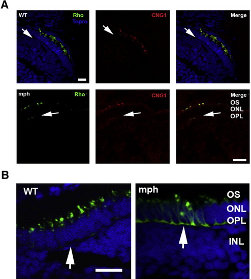
Mis-localization of the photopigment apoprotein in cones and to a lesser extent in rods of morphants. (A) (Upper panel) Double label of rhodopsin (green) and CNG1 (red) in rods of 3-day-old control (WT) and morphants. Topro (blue) was used in the control (WT) to show the nuclear layers. Note that Rho and CNG1 co-localize in the OS in control (WT) and are undetectable in the outer plexiform layer (arrows). (Lower panel) In contrast to control (WT), both Rho and CNG1 are seen in the outer plexiform (synaptic) layer of morphants (arrows). Bar = 20.55 μm. (B) Cone opsin (green) is very extensively mis-localized to the perinuclear region and outer plexiform layer (synaptic layer) in morphant (mph) cones compared to control (see arrows). Sections are double labeled with Topro (blue) to show the nuclear layers. Bar = 12.5 μm. |
| Gene: | |
|---|---|
| Fish: | |
| Knockdown Reagent: | |
| Anatomical Terms: | |
| Stage: | Protruding-mouth |
| Fish: | |
|---|---|
| Knockdown Reagent: | |
| Observed In: | |
| Stage: | Protruding-mouth |
Reprinted from Developmental Biology, 316(1), Insinna, C., Pathak, N., Perkins, B., Drummond, I., and Besharse, J.C., The homodimeric kinesin, Kif17, is essential for vertebrate photoreceptor sensory outer segment development, 160-170, Copyright (2008) with permission from Elsevier. Full text @ Dev. Biol.