
Fig. S1
- ID
- ZDB-FIG-080327-3
- Publication
- Sun et al., 2008 - Genome-Wide Survey and Developmental Expression Mapping of Zebrafish SET Domain-Containing Genes
- Other Figures
- All Figure Page
- Back to All Figure Page
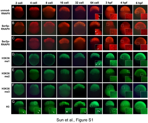
Immunofluorescent analyses of RNA polymerase II phosphorylation and histone H3K36 methylation in zebrafish embryos. Zebrafish embryos at different stages were subject to immunofluorescent staining to detect the unmodified pol II (A) and hyperphosphorylated pol II (B and C), H3K36 monomethylation (D), dimethylation (E) and trimethylation (F). Immunofluorescent staining of histone H3 (G) was used as a positive control. While the staining of histone H3 in nuclei is consistently detected (G), the staining of H3K36 methylation cannot be detected until 64-cell stage (D–F). The inset panels show the magnified views of detected staining in nuclei (arrow head). The unmodified, serine 2-phosphorylated and serine 5-phosphorylated pol II were probed with mouse monoclonal antibodies 8WG16, H5 and H14 (Covance Research Products), respectively. H3K36 mono-, di- and trimethylation were probed with rabbit polyclonal antibodies ab9084 (ABcam), 07-274 (Upstate) and ab9050 (Abcam), respectively. Histone H3 was probed with rabbit polyclonal antibody ab1791 (Abcam). |