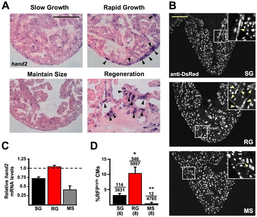
Cardiogenic markers are induced during cardiac homeostasis. (A) hand2 in situ hybridization of ventricular sections. hand2 is weakly expressed in MS hearts, mildly expressed in SG hearts and more strongly expressed in RG hearts similar to regenerating hearts 7 days post-resection (arrowheads show foci of strong expression). (B) Sections from SG, RG and MS ventricles of cmlc2:nRFP zebrafish, stained for DsRed immunoreactivity. RG ventricles show many more RFPcyto cells, a marker that suggests recent CM differentiation events (arrowheads in insets). (C) Real-time Q-PCR analysis of hand2 expression from whole ventricles of SG, RG and MS hearts, relative to expression in ventricles 7 days after apical resection. hand2 expression is similar in ventricles from RG fish to that in regenerating ventricles (1.0, represented by dashed line) and greater than in SG and MS ventricles. (D) Quantification of RFPcyto cells in SG, RG and MS groups. The average RFPcyto index per animal is plotted. Numbers above the error bars indicate RFPcyto CMs over the total CMs counted from all animals combined. Numbers below group abbreviations indicate the number of animals/ventricles per group (*P<0.01, t-test, significantly different from SG; **P<0.005, t-test, significantly different from SG and RG). Scale bars: 100 μm.
|
