
Fig. S6
- ID
- ZDB-FIG-080325-87
- Publication
- Bessa et al., 2008 - meis1 regulates cyclin D1 and c-myc expression, and controls the proliferation of the multipotent cells in the early developing zebrafish eye
- Other Figures
- All Figure Page
- Back to All Figure Page
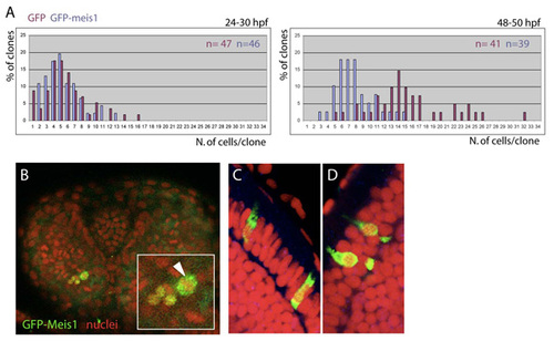
Size of GFP-meis clones at two developmental stages and subcellular localization of the GFP-Meis product. (A) Size of GFP and GFP-meis1 clones at two developmental stages. The number of clones (n) for each clone type is indicated. (B) 30-hpf eye containing GFP-meis1-expressing cells counterstained with DAPI to label DNA. GFP-meis1 cells can be seen undergoing mitosis (arrowhead in the inset). (C,D) 3-dpf GFP-meis1-expressing clones can only be recovered in the photoreceptor layer. Here, the GFP-tagged Meis1 transcription factor accumulates mostly in the cytoplasm of the photoreceptors. This contrasts with its nuclear accumulation at earlier stages (see Fig. 4). The nuclear localization of Meis proteins have been shown to depend on their interaction with Pbx products (Vlachakis et al., 2001; Choe et al., 2002). Therefore, one possible cause for the cytoplasmic accumulation of GFP-Meis1 in photoreceptors might be reduced levels, or absence, of Pbx partners in these cells. In addition, Pbx proteins can be retained in the cytoplasm by direct binding to non-muscle myosin II heavy chain B (Huang et al., 2003). If this were the case in photoreceptor cells, the Meis1-Pbx complex would not be able to travel to the nucleus. Alternatively, the overexpressed GFP-Meis1 might be saturating the nuclear import machinery, causing a large fraction of this protein to remain in the cytoplasm. |