
Fig. 6
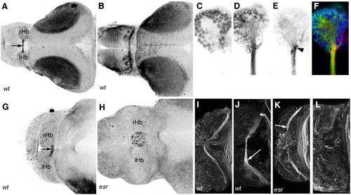
EphB receptors are surface-localized at the roof plate boundary. (A) Ephrin-B2/Fc binds to HC axons only within the commissure (arrow) at 3 dpf. (B) Ephrin-A2/Fc affinity probe indicates surface localization of EphA receptors in the HC, the habenular neuropils, stria medullaris and tract of the habenular commissure at 3 dpf. (C–F) Higher magnification view of the right habenula labeled with the nuclear stain Syto-11 (C), acetylated tubulin (D) and Ephrin-B2/Fc (E) shows that EphB receptors are surface-localized at the medial boundary of the habenula (arrowhead). (G, H) Ephrin-B2/Fc binding is detected in a wild type (G) but not in a mutant (H) at 2 dpf. (I–L) Immunofluorescence with a pan EphB antibody at 2 (I, K) and 3 (J, L) dpf. Labeling is detected in wild type habenular commissure axons, with stronger label in the commissure at 3 dpf (I, arrow). Mutant axons are labeled at 2 dpf (K, arrow), but not at 3 dpf (L). rHb: right habenula; lHb: left habenula. |
Reprinted from Molecular and cellular neurosciences, 37(2), Hendricks, M., Mathuru, A.S., Wang, H., Silander, O., Kee, M.Z., and Jesuthasan, S., Disruption of Esrom and Ryk identifies the roof plate boundary as an intermediate target for commissure formation, 271-283, Copyright (2008) with permission from Elsevier. Full text @ Mol. Cell Neurosci.