
Fig. 5
- ID
- ZDB-FIG-080225-24
- Publication
- Patterson et al., 2008 - Growth in the larval zebrafish pectoral fin and trunk musculature
- Other Figures
- All Figure Page
- Back to All Figure Page
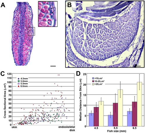
Addition of new muscle fibers occurs at the periphery of the existing muscle masses. A: Transverse section of the pectoral fin of a 6.5 mm larva stained with methylene blue and basic fuchsin. Fibers with a small cross-sectional area (CSA) are closest to the skin. Inset shows small fibers near the skin. B: Methylene blue-stained transverse section through an adductor muscle in the pectoral fin of an adult (32 mm). No small, new fibers are within the mass of the muscle. Ventral is to the bottom. C: Scatter plot showing the relationship between the CSA of a muscle fiber and its position relative to the skin and endoskeletal disk. Each point represents one muscle fiber. Distances were determined using the GIS program ArcGIS by mapping the relationship between the center of each fiber and the closest point on the skin or endoskeletal disk. D: Chart showing the relationship between bins of fiber size (CSA) and the median distance from the skin. Muscle fibers that are less than 10 μm2 in CSA have a smaller median distance from the skin in all larval specimen sizes (4.5 mm, n = 3; 5.5 mm, n = 3; 6.5 mm, n = 4). Scale bar = 25 μm in A, 100μm in B. |