
Fig. 6
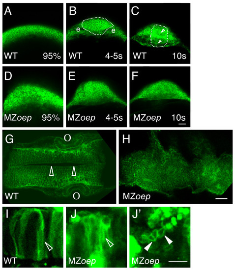
The ordered structure of the neural tube is disrupted in MZoep embryos. (A-H) Embryos were injected at the one cell stage with mGFP mRNA and imaged (A-F) live at 95% epiboly (95%), the 4–5 somite stage (4–5 s), the 10 somite stage (10 s), or (G, H) fixed at ~24 hpf. (A-F) Cross sections through the anterior developing neural plate, with presumptive eyes (e) indicated in panel B, the outer boundary of the neural tube indicated by a dotted line in panels B and C, and the midline indicated by the open arrowheads in C. (G, H) Horizontal sections through the midbrain and hindbrain of ~24 hpf embryos, with the midline of the brain (open arrowheads) and the otic vesicles (o) indicated in panel G. (I-J′) Embryos were injected at the one cell stage with DNA encoding mGFP, raised to ~24 hpf, and imaged live in high magnification horizontal sections. Open arrowheads indicate elongated cells and the closed arrowheads indicate round cells. All images are confocal optical sections. Scale bars: 40 μm (A-H), 80 μm (I-J′). |