
FIGURE
Fig. 4
- ID
- ZDB-FIG-080117-3
- Publication
- Godinho et al., 2007 - Nonapical symmetric divisions underlie horizontal cell layer formation in the developing retina in vivo
- Other Figures
- All Figure Page
- Back to All Figure Page
Fig. 4
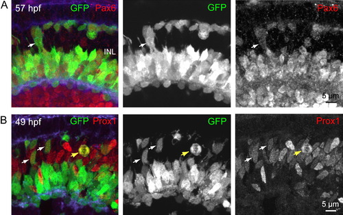
HC Precursors Express Progenitor Markers (A) Immunostaining of a retinal cross-section from a ptf1a:GFP zebrafish for Pax6, a progenitor cell marker. Arrow points to an example of an HC precursor-like GFP+ cell that expressed Pax6. (B) Immunolabeling for Prox1 demonstrates that it is expressed in GFP+ cells during M phase (yellow arrow) of the cell cycle. Examples of HC precursor-like GFP+ cells that express Prox1 (49 hpf) are indicated by white arrows. INL, inner nuclear layer. |
Expression Data
| Genes: | |
|---|---|
| Fish: | |
| Anatomical Term: | |
| Stage: | Long-pec |
Expression Detail
Antibody Labeling
Phenotype Data
Phenotype Detail
Acknowledgments
This image is the copyrighted work of the attributed author or publisher, and
ZFIN has permission only to display this image to its users.
Additional permissions should be obtained from the applicable author or publisher of the image.
Full text @ Neuron