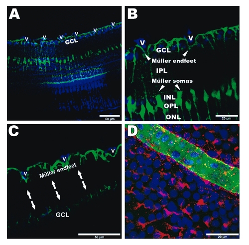
Physical interaction of Müller glia and retinal vasculature in adult zebrafish. A: Transverse view of peripheral retina in an adult Tg(gfap:EGFP) transgenic animal shows Müller cells (green) expanding through all retinal layers (blue: nuclear DAPI staining). B: Higher magnification shows Müller endfeet interposed with ganglion cell soma and contacting the retinal vessels (v). C: When the vascular layer is dissected from the inner interface of the retina (arrows) Müller endfeet remain attached to the vessels indicating a tight interaction. D: Blood vessel (green: Fli1-EGFP) overlying an adult retina seen from above with ganglion cell layer in the background (blue: DAPI nuclear staining). Müller cell endfeet (red: GFAP antibody) are observed on the entire surface of the inner retina, but especially concentrated along the retinal vessel, in direct contact with the vascular endothelium (yellow co-staining). GCL: ganglion cell layer; v: vessel; IPL: inner plexiform layer; INL: inner nuclear layer; OPL: outer plexiform layer; ONL: outer nuclear layer.
|
